
 ChatGPT

Medically reviewed by Prof. Dr. Özgür Pilancı, Plastic, Reconstructive & Aesthetic Surgeon — August 2026
Quick answer (updated August 2026): The nose has three structural thirds — bone at the top, cartilage in the middle, and a flexible cartilage-and-skin tip at the bottom — and almost every term in a rhinoplasty consultation refers to a landmark in one of them. The alar is the curved outer wall of each nostril. The columella is the strip of tissue between the nostrils. The dorsum is the bridge. The nares are the nostril openings themselves. This glossary defines every term you are likely to meet, in plain English, and explains what each one means when it appears in a surgical plan.
How the nose is built
Before the vocabulary, the architecture. The nose is three stacked units, and knowing which one a term belongs to makes the rest easier.
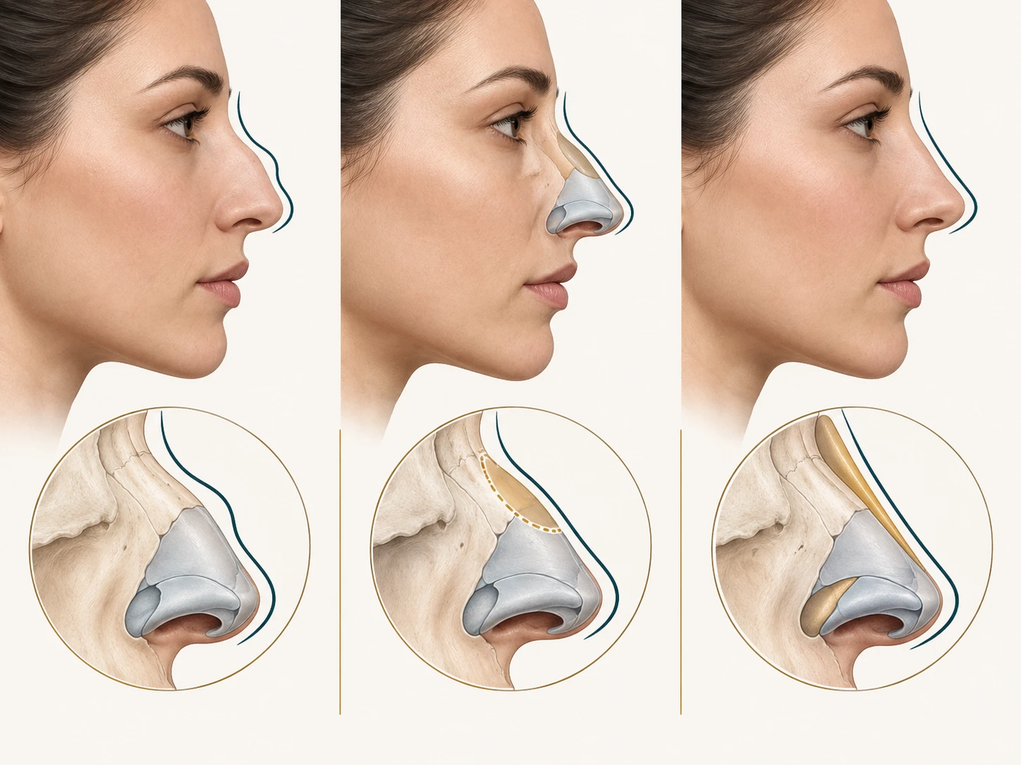
The upper third is bone: the paired nasal bones, meeting the frontal bone at the top and the maxilla on each side. This is the rigid part, and the only part that requires controlled bone cuts to move.
The middle third is the cartilaginous vault: the paired upper lateral cartilages fused to the top edge of the septum. It is springy rather than rigid, and it forms the roof of the nose’s most important breathing space.
The lower third is the tip: the paired lower lateral cartilages, each shaped like a curved strip with three sections — the medial crus running up the columella, the middle (intermediate) crus forming the dome at the tip, and the lateral crus curving out into the nostril wall. This unit is mobile, and it is where most of the fine work in rhinoplasty happens.
Where the bone and cartilage meet is the keystone area, the junction of the nasal bones, upper lateral cartilages and septum. It is the nose’s central support, and preserving it is the entire idea behind preservation rhinoplasty.
Surface landmarks: the outside of the nose
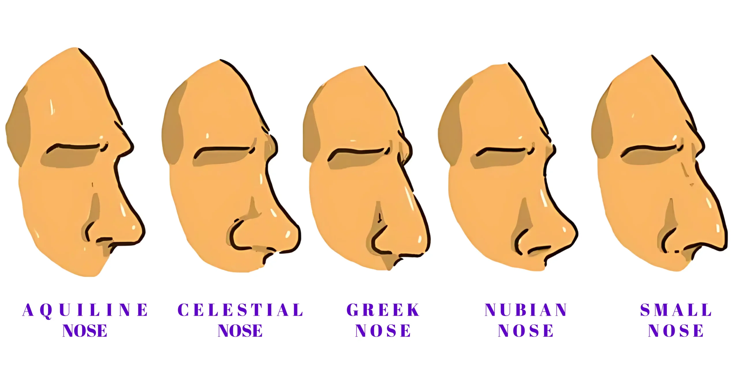
Radix (nasion). The root of the nose — the deepest point of the curve between the forehead and the bridge, roughly level with the eyelid crease. Its depth and height set where the nose appears to "start", which is why raising or lowering the radix changes apparent nose length without touching the tip.
Rhinion. The point where the nasal bones end and cartilage begins. The skin is at its thinnest here, which is why irregularities along the bridge show up at this exact spot.
Dorsum (the nasal bridge). The ridge running from radix to tip. Bone above, cartilage below. A dorsal hump is excess height along it; a low or flat nasal bridge is a dorsum sitting at or below the level of the eyes in profile; a high nasal bridge is one with a pronounced projecting line. None of these is a defect — they are different structures needing different work: reduction for one, augmentation with grafts for the other.
Supratip break. The subtle shadow just above the tip where the dorsal line steps down before rising into the tip itself. It is a small feature that does a lot of work: a nose without it looks heavy at the tip, and its loss after surgery is the defining sign of a polly beak deformity.
Tip-defining points. The two highest, most forward points of the tip — one on each dome. In a photograph they appear as two small highlights, and creating a clean pair of them is a large part of what tip surgery is trying to achieve.
Infratip lobule. The small area between the tip-defining points and the point where the columella begins.
Columella. The strip of skin and cartilage running between the nostrils, from the tip down to the upper lip. It houses the medial crura. Columellar show is how much of it is visible in profile — normally 2 to 4 mm. More than that is a hanging columella; less than that is a hidden or retracted columella.
Nasolabial angle. The angle between the columella and the upper lip — the measure of how "upturned" a nose looks. Commonly cited aesthetic ranges are around 90–95° in men and 95–105° in women. A retroussé nose is simply an upturned one, with a wider angle.
Nasofrontal angle. The angle at the radix, between the forehead and the bridge, typically cited around 115–130°.
Ala (plural: alae). The curved, fleshy outer wall of each nostril — the "wing" of the nose, which is what the Latin means. Supported internally by the lateral crus. This is the single most-searched anatomical term on the nose, and almost everything with "alar" in it refers back to it.
Alar rim. The free curved edge of the ala, forming the outer boundary of the nostril opening.
Alar base. Where the ala meets the cheek and upper lip at the bottom of the nostril. Alar base width — the distance between the two — is the measurement behind "wide nostrils".
Alar crease / alar-facial groove. The natural crease where the ala meets the cheek. Incisions for alar base reduction are placed inside it precisely because the resulting scar disappears into a line that already exists.
Nasal sill. The floor of the nostril opening — the horizontal band of tissue at the base of each nostril, running from the columella outward to the alar base.
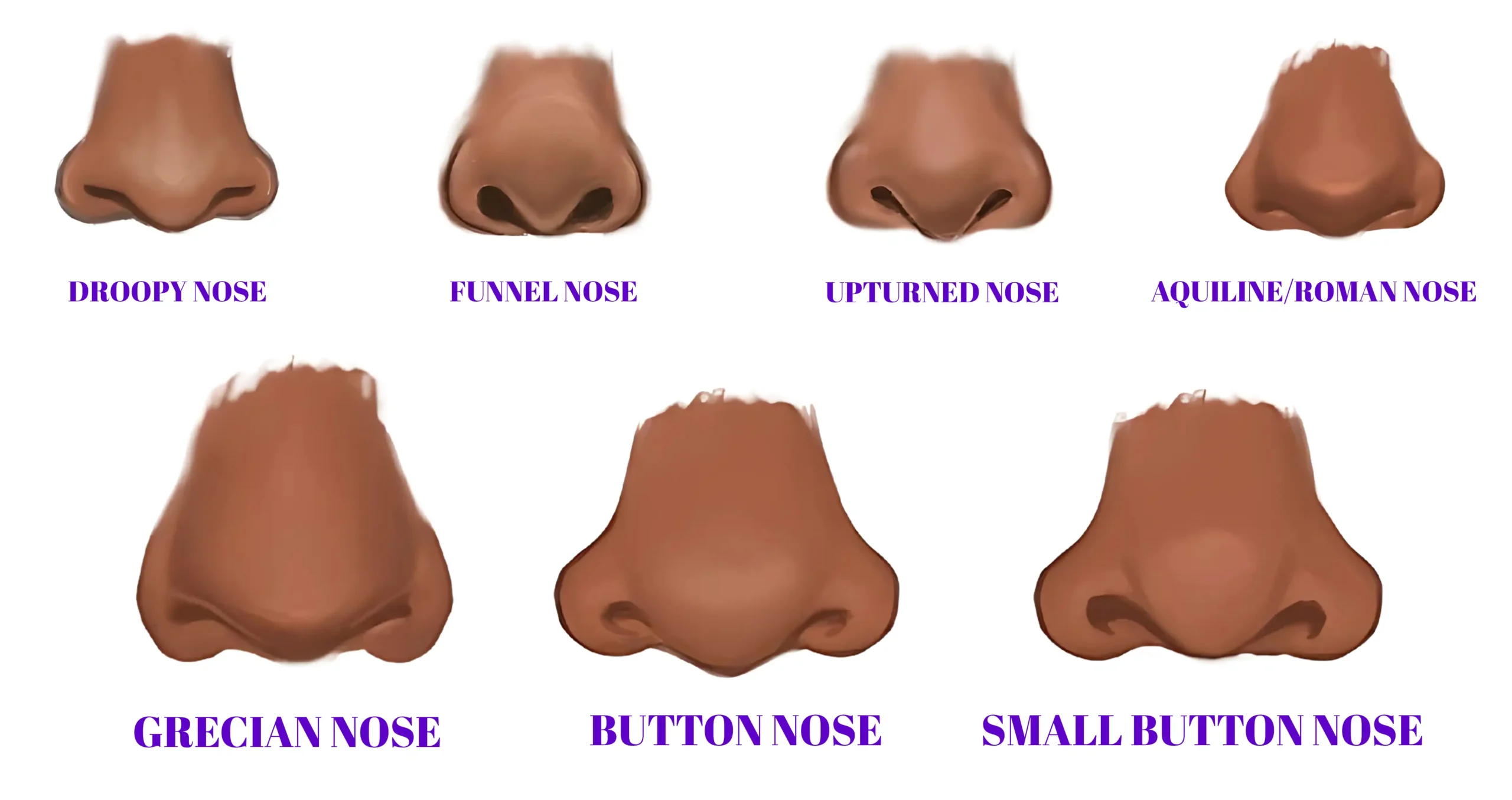
Nares. The nostril openings themselves. "What are nares" is one of the most common searches on this list, and the answer is simply: the two external openings of the nose. Their shape in base view — pear-shaped, oval, round — varies enormously and is part of what makes a nose recognisable.
Soft triangle (facet). The small area of folded skin just inside the alar rim, at the top of each nostril, containing no cartilage at all. It is delicate, it heals badly, and a notch in it is one of the more visible signs of a rhinoplasty.
Footplates. The flared lower ends of the medial crura, at the base of the columella. They are part of the tip’s support structure, and prominent footplates can widen the base of the columella visibly.
Inside the nose
Septum. The central wall dividing the two nasal passages. Cartilaginous at the front — the quadrangular cartilage — and bony at the back, formed by the perpendicular plate of the ethmoid above and the vomer below. It is both a divider and a structural pillar; over-harvesting it for grafts is one route to a collapsed bridge.
Membranous septum. The short, cartilage-free segment between the front edge of the septal cartilage and the columella. Its flexibility is why the tip can move.
Internal nasal valve. The narrowest part of the entire airway, formed where the upper lateral cartilage meets the septum, with a normal angle of about 10–15°. Because it is the narrowest point, it is the flow-limiting one — which is why a few degrees lost here causes a disproportionate sense of blockage. Collapse follows over-resection of the bridge, trauma, or age-related cartilage weakening, and is the reason spreader grafts exist.
External nasal valve. The nostril opening itself, bounded by the alar rim, the columella and the sill. Collapse here is what makes the nostril wall suck inward on hard inspiration.
Turbinates. The scroll-shaped shelves on the outer wall of each nasal passage — inferior, middle and superior — that warm and humidify air. Enlarged inferior turbinates are a very common cause of blockage, and turbinate reduction often accompanies septal surgery.
Sesamoid (minor alar) cartilages. Small accessory cartilages linking the lateral crus to the bony rim of the nasal opening, contributing to sidewall support.
The muscles
Nasalis. The largest nasal muscle, in two parts with opposite jobs: the transverse part compresses the nostrils, and the alar part widens them. It is why nostrils flare with effort or emotion.
Procerus. The muscle between the brows that pulls the skin over the radix downward, producing the horizontal lines across the top of the nose.
Levator labii superioris alaeque nasi. The long name belongs to the muscle that lifts both the upper lip and the ala — the one that produces the "sneer".
Depressor septi nasi. A small muscle running from the upper lip to the base of the septum. When it contracts on smiling, it pulls the tip downward. If your nose visibly droops when you smile, this muscle is usually the reason, and releasing it is a routine part of many tip procedures.
Terms for problems, and what they actually mean
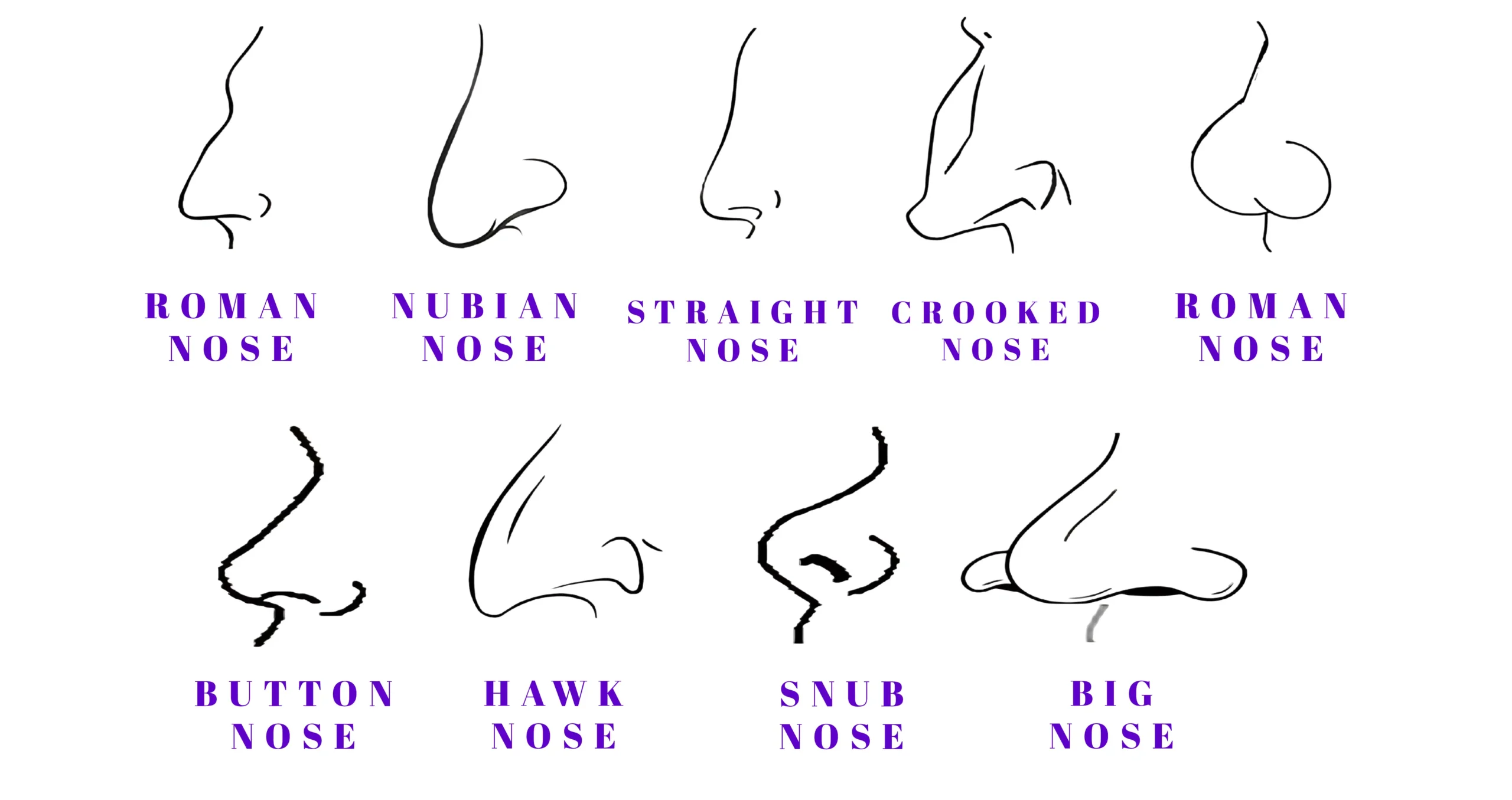
Dorsal hump. Excess bone and cartilage along the bridge. Reduced by rasping and trimming, which leaves an "open roof" that lateral osteotomies then close.
Bulbous nasal tip. A round, undefined tip. Two causes, often together: thick tip skin masking the underlying shape, and lower lateral cartilages that are wide or convex. Addressed with cartilage trimming and suturing — with the honest caveat that thick skin limits how much definition is achievable, because the definition has to show through it.
Bifid nasal tip. A visible cleft or double point at the tip, caused by the two domes sitting too far apart. Corrected by suturing them closer together.
Boxy tip. A squared-off tip, from a wide angle between the domes or a broad dome arc.
Hanging columella. Columellar show greater than about 4 mm, so the columella hangs below the alar rims in profile. Usually excess caudal septum or over-long medial crura; corrected by trimming or suture repositioning.
Alar retraction. The opposite problem — the alar rim sits too high, exposing more nostril than it should. Usually caused by over-resection of the lateral crura in previous surgery, or by scar contracture. It is one of the harder things to correct, requiring rim, batten or composite grafts.
Alar flaring versus a wide alar base. These are different things and get confused constantly. Flaring is outward convexity of the ala beyond a vertical line dropped at the alar-facial groove — the nostrils bow outward. A wide base means the distance between the two alar bases exceeds the distance between the inner corners of the eyes. They need different operations: flare is reduced with a wedge excision at the alar crease, width with a sill excision. A surgeon who treats the two identically will over-narrow one and under-correct the other.
Polly beak. Fullness in the supratip area that erases the supratip break, leaving a parrot-beak profile. The cartilaginous version comes from leaving too much dorsal septal cartilage; the soft-tissue version comes from scar filling a space that was over-resected — more common in thick skin, and much harder to fix.
Inverted-V deformity. A visible step-off where the nasal bones meet the upper lateral cartilages, appearing as an inverted V across the middle of the nose. It follows hump removal without restoring middle-vault support, and it is the classic argument for spreader grafts.
Saddle nose. A collapsed, concave bridge from loss of septal support — after trauma, infection, or over-aggressive surgery. Corrected by structural augmentation.
Ptotic (drooping) tip and over-rotated tip. Tip rotation describes where the tip points. Too far down reads as heavy and ageing; too far up reads as artificial and exposes the nostrils. Both are adjusted with grafts and sutures rather than by removing tissue.
The measurements used in planning
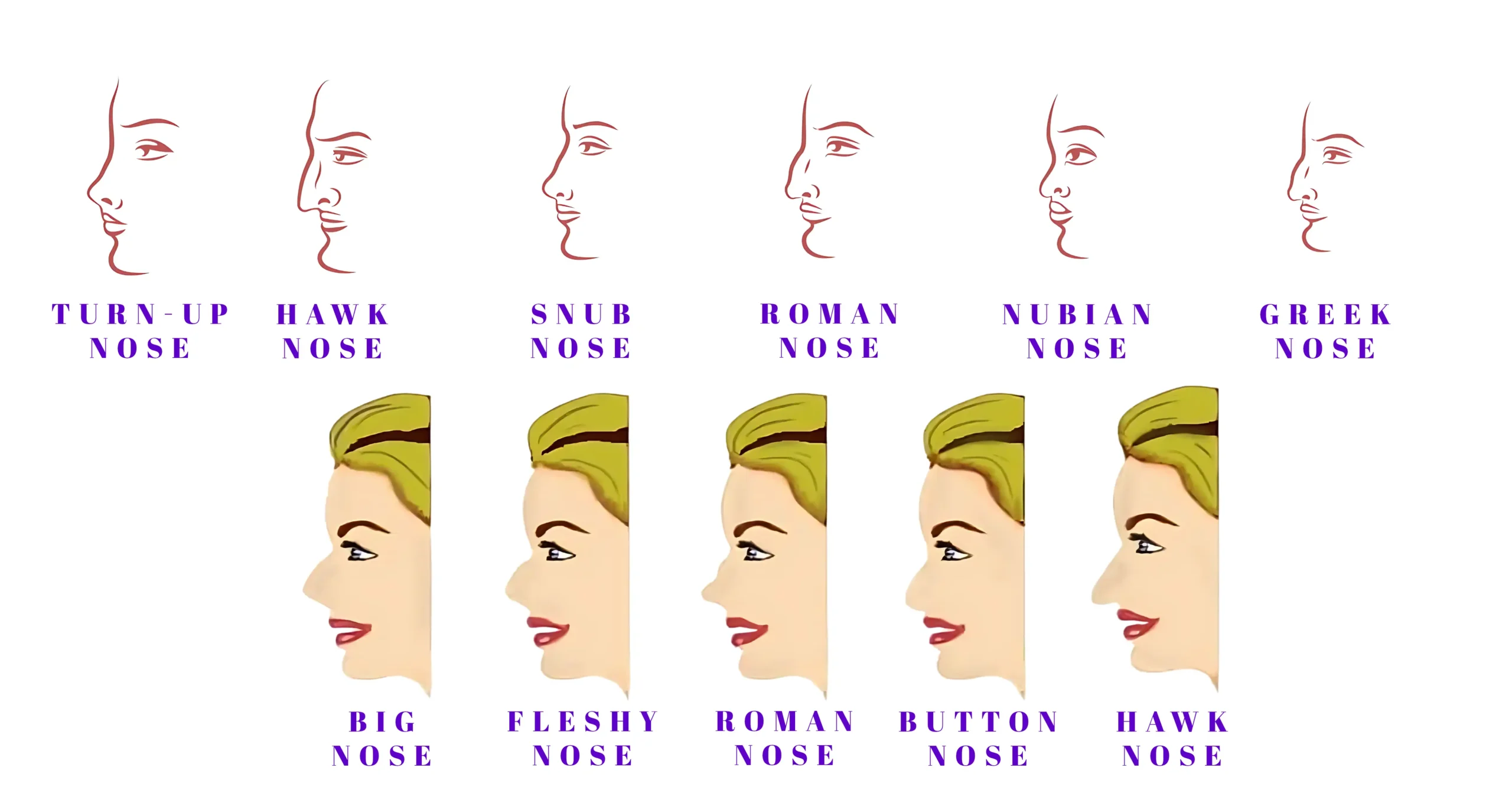
Rhinoplasty planning uses a handful of reference proportions. They are averages drawn from aesthetic-preference studies — many of them conducted on predominantly European faces — and they exist to help a surgeon think about balance, not to define a correct nose. A nose that suits a face is the goal; a nose that hits a number is not.
- Interalar width compared with intercanthal distance — the width across the alar bases against the distance between the inner corners of the eyes.
- Tip projection, expressed as Goode’s ratio, commonly cited at around 0.55–0.60.
- Nasolabial angle, around 90–95° in men and 95–105° in women.
- Columellar show, 2–4 mm.
- Nasofrontal angle, around 115–130°.

Skin thickness: the variable nobody mentions
Nasal skin is not uniform. It is thinnest over the rhinion — the middle of the bridge — and thickest and most sebaceous over the tip and supratip.
This matters more than most patients are told. Thick tip skin hides the cartilage work underneath it, limits how much definition is achievable, and raises the risk of soft-tissue polly beak because scar tissue fills the space where cartilage was removed. Thin skin shows everything, including graft edges and tiny irregularities that thicker skin would conceal. Neither is better; each changes what the operation can promise, and a surgeon who assesses your skin thickness and tells you what it means is giving you the most useful information in the consultation.
Terms you may see in a surgical quote

Open versus closed approach. Closed (endonasal) rhinoplasty works entirely through incisions inside the nostrils. Open rhinoplasty adds a small incision across the columella, lifting the skin to expose the framework directly — more visibility, a small columellar scar, generally preferred for complex, revision and thick-skinned cases.
Osteotomy. A controlled cut in the nasal bones. Lateral osteotomies narrow the nose and close the open roof left by hump removal; medial osteotomies mobilise the bones from the centre.
Spreader graft. A strip of cartilage placed between the septum and the upper lateral cartilage to hold the internal valve open and prevent an inverted-V deformity. One of the most common grafts in modern rhinoplasty, and largely why airway function survives cosmetic bridge reduction.
Columellar strut. A cartilage graft between the medial crura, supporting tip projection.
Tip or shield graft. Cartilage placed over the domes to increase projection and refine the infratip lobule.
Cephalic trim. Removing the upper portion of the lateral crus to narrow a bulbous tip — with a preserved rim of cartilage, conventionally around 6–8 mm, left behind. Take too little and the tip stays round; take too much and the nostril rim collapses or retracts years later.
Alar base reduction (Weir excision). Wedge excisions at the alar base to reduce flare, width, or both.
Preservation rhinoplasty. An approach that lowers the dorsum as a unit rather than removing the hump and rebuilding, keeping the keystone area and the dorsal ligaments intact. It suits some noses very well and others not at all.
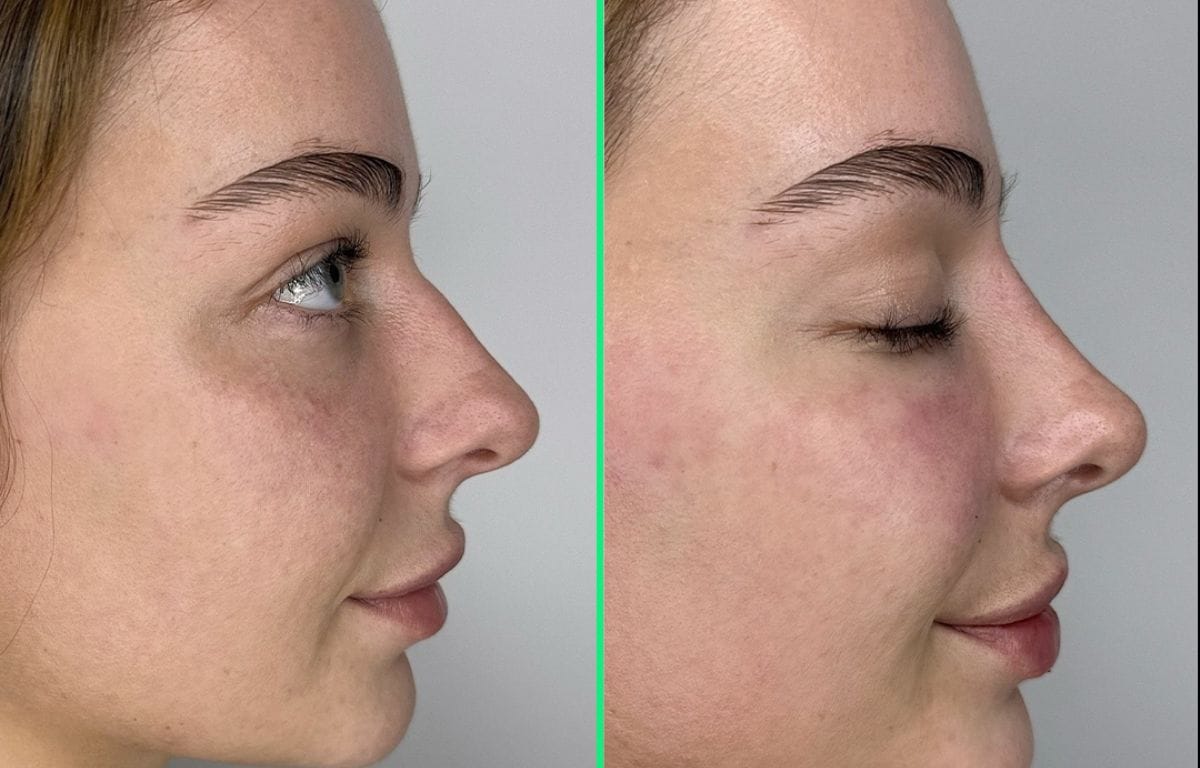
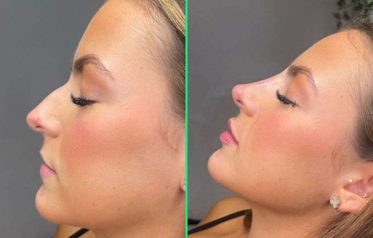
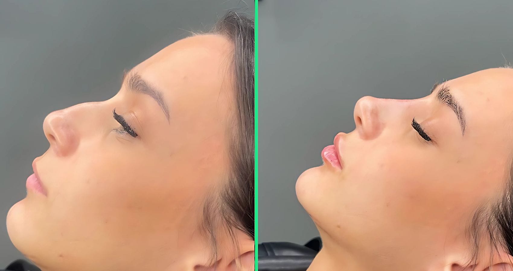
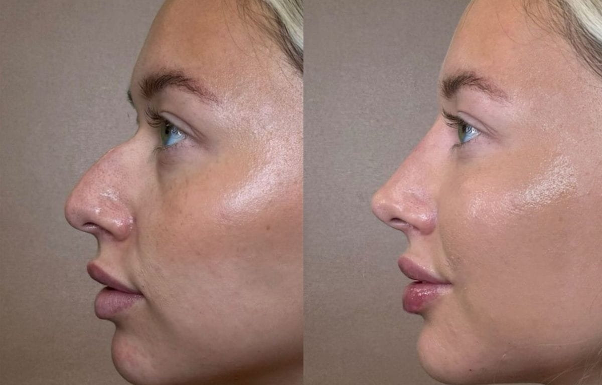
You can see how these decisions read on real faces in our before-and-after gallery, and our rhinoplasty page covers the operation itself.
Rhinoplasty Before & After Results at Vivid Clinic



Frequently Asked Questions
What does "alar" mean?
Alar refers to the ala — the curved, fleshy outer wall of each nostril, from the Latin for "wing". Almost every compound term follows from it: the alar rim is its free edge, the alar base is where it meets the cheek and lip, the alar crease is the groove between the two, alar flaring means the wall bows outward beyond the base, and alar retraction means it sits too high and shows too much nostril.
What are nares?
Nares are the nostrils — specifically the two external openings of the nose. Their shape in base view varies widely between individuals and is one of the features that makes a nose distinctive. The singular is naris.
Where is the columella and how much should show?
The columella is the strip of skin and cartilage running between the two nostrils, from the tip of the nose down to the upper lip. In profile, 2 to 4 millimetres of it should be visible below the alar rims. More than about 4 mm is described as a hanging columella; less than 2 mm as a retracted or hidden one. Both are adjustable, usually by reshaping the caudal septum or repositioning the medial crura.
What is the difference between a high and a low nasal bridge?
The bridge, or dorsum, is the ridge running from between the eyes to the tip. A high bridge projects forward with a pronounced profile line; a low or flat bridge sits closer to the level of the eyes with a shallower profile. Neither is a defect — they are different structures common in different populations, and they need opposite work: a high bridge with an unwanted hump is reduced, while a low bridge is augmented with cartilage grafts or, in some practices, an implant.
What causes a bulbous nasal tip?
Two things, usually together: thick, sebaceous skin over the tip that masks the shape beneath it, and lower lateral cartilages that are wide or convex rather than neatly domed. Surgery narrows and reshapes the cartilage with trimming and sutures, but skin thickness sets the ceiling on how much refinement will actually be visible — which is why an honest surgeon discusses your skin before promising a defined tip.
Which nose measurements actually matter?
The commonly cited ones are interalar width against intercanthal distance, tip projection (Goode’s ratio around 0.55–0.60), nasolabial angle (about 90–95° in men and 95–105° in women), and columellar show of 2–4 mm. They are aesthetic averages, many derived from studies of predominantly European faces, and they are planning references rather than targets. Balance with the rest of your face — and your airway — outrank any of them.
Met a term in a quote that nobody explained? A good consultation should leave you able to describe your own operation in your own words. Send photographs for a free online assessment and get a plain-English explanation of exactly which structures would change, and what each change would do.